OCT-SD-01
The ability to image samples with high resolution makes it suitable for applications where small areas are examined in detail.
Example of OCT-SD-01 imaging①
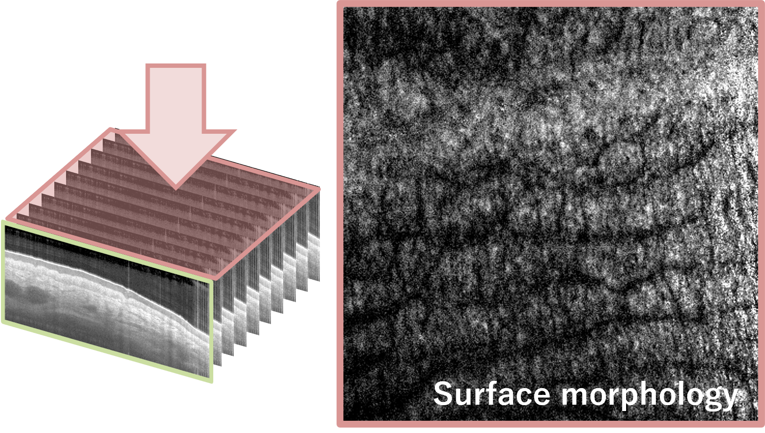
Below is the human finger measured by OCT-SD-01.
A surface image and multiple tomographic (cross-sectional) images can be obtained from the 3D data measured after a few seconds of scanning.
In the surface image, surface morphology such as fine wrinkles, texture, and pores could be confirmed.
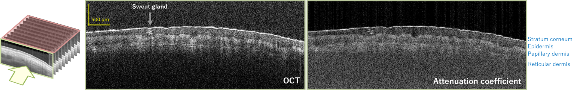
In the tomographic image, the layer structure and sweat glands near the surface could be clearly observed.
We post new imaging examples on our Facebook page and Twitter from time to time.
Example of OCT-SD-01 imaging②
The figure below shows the measurement of a cucumber slice.
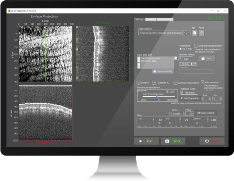
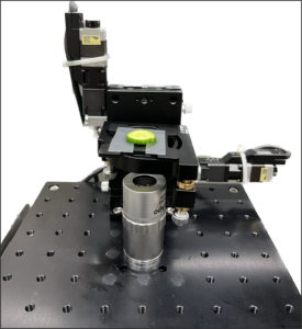
In OCT-SD-01, the sample is placed on a motorized stage to facilitate fine adjustment of its position. In addition, optical microscope images of the OCT measurement area can be acquired with a CCD camera.
These allow the motorized stage to accurately set the desired measurement point while observing with a CCD camera.
In this case, we targeted the area around the embryo (radicle) portion of the cucumber seed and took measurements.
The figure below shows an optical microscope image (left) and an OCT cross-section image (upper right) of the red line area in the optical microscope image.
Because the sample is thick, the cell wall cannot be observed with an optical microscope (left), but with OCT (right), the cell wall of the cucumber is clearly visible. OCT is characterized by its ability to acquire cross-section images of thick samples cut in depth.
OCT also provides an image cut at each depth (Enface image). The Enface image at the depth of the green line in the OCT cross-section image (upper right) is in the lower right of the figure below. It looks like a microscope image.
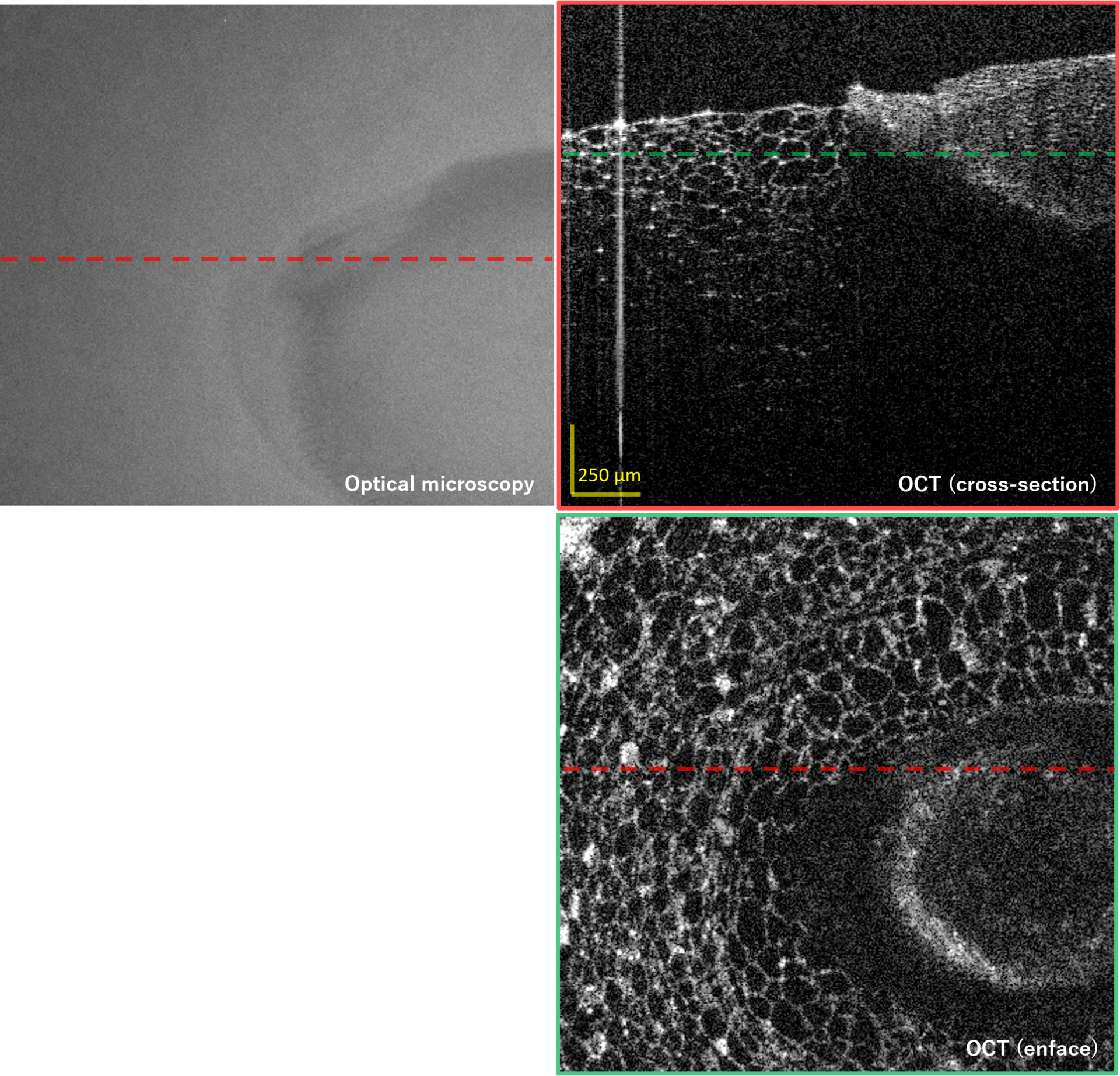
Optical microscope image and OCT images of the cucumber slice
With OCT, 3D data can be acquired in a few seconds of scanning. See the data in the video below. The cell walls look very beautiful.
Movie of enface images (horizontal cross-section)
Movie of enface images (vertical cross-section)
Sample images of OCT-SD-01
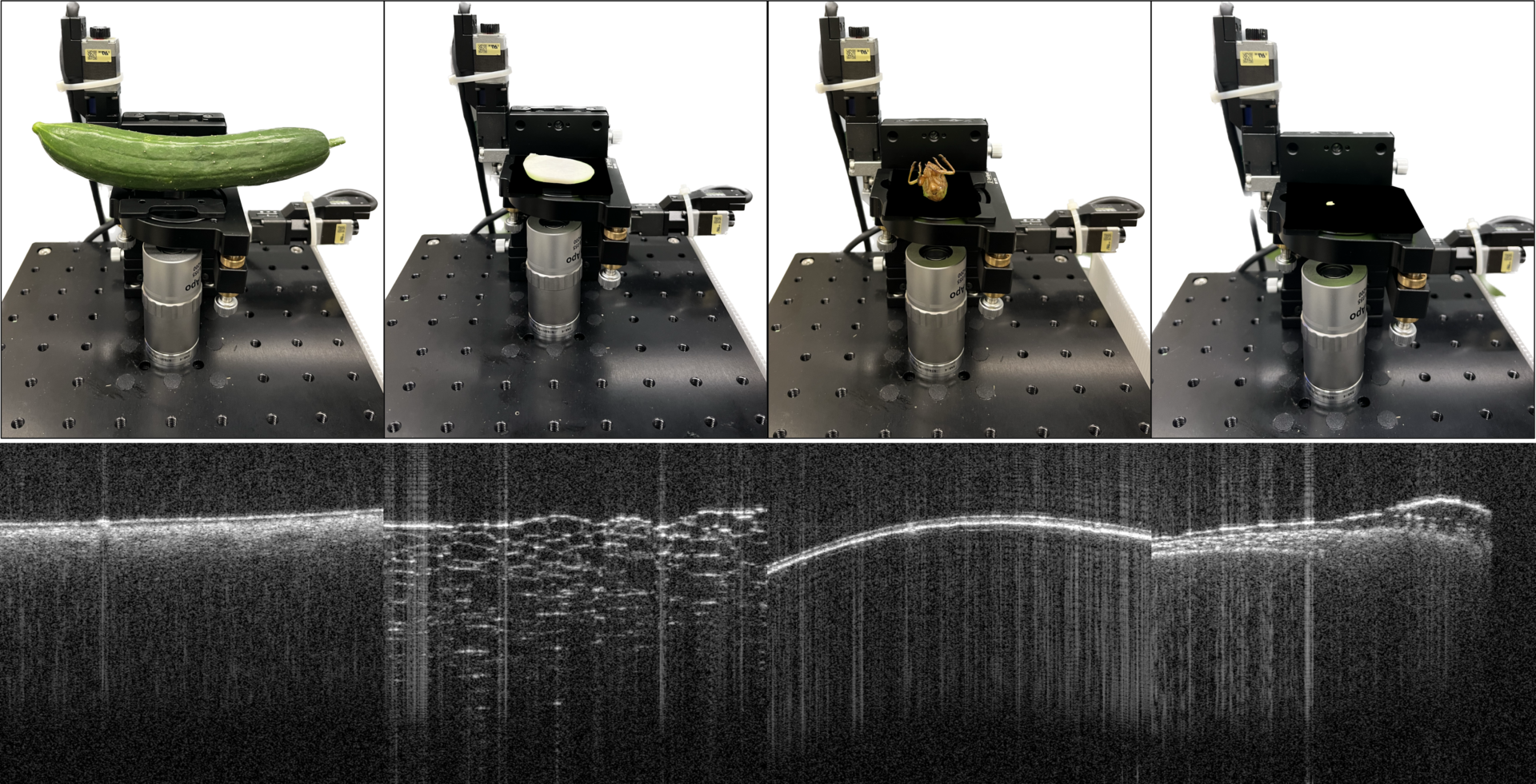
Cucumber, radish slice, cicada shell, and green pepper seed
Upper: Photo
Lower: OCT cross-section image
OCT-SD-01 specifications
| Lateral resolution | Depth resolution | Imaging depth | Scan range | A-scan rate | B-scan rate | Volume scan time |
| 15 μm | 10 μm (in air) 7.3 μm (in tissue) | 1 mm (depends on the sample) | 4 × 4 × 4 mm3 (you can select 1-12 mm) | 50,000/sec | 110/sec | 4.7 sec |
Functions and performance can be customized according to the measurement target and application. For details, please contact us.
OCT product specs comparison chart