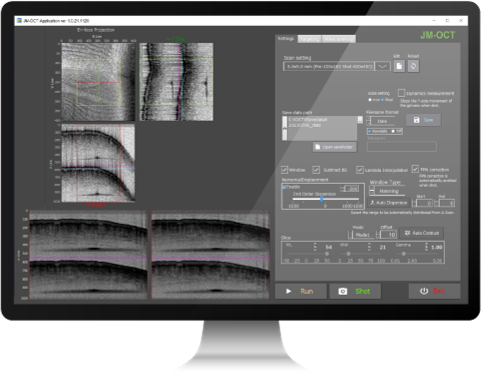
OCT-JM-01
The ability to measure birefringence makes samples can be analyzed using multiple optical properties. It expands the scope of research.
In addition to industrial applications, this technology is also attracting attention as an evaluation technique for cultured tissues used in drug discovery, regenerative medicine, and biological research. It is expected to be used in a wide range of fields.
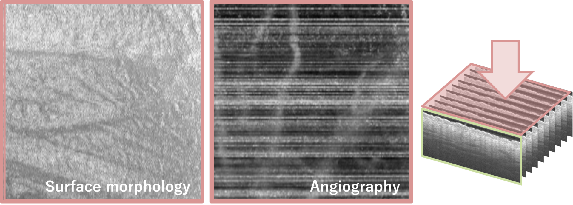
Example of OCT-JM-01 imaging ①
Below is the human finger measured by OCT-JM-01.
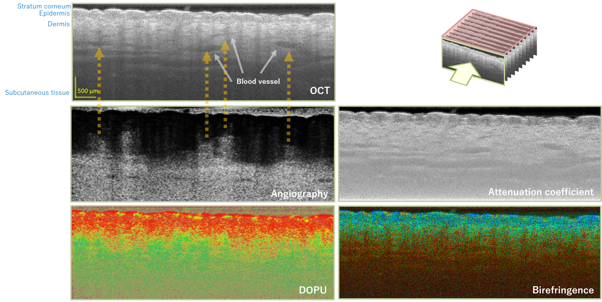
From the Jones Matrix 3D data measured after a few seconds of scanning, a surface image and multiple tomographic (cross-sectional) images can be obtained.
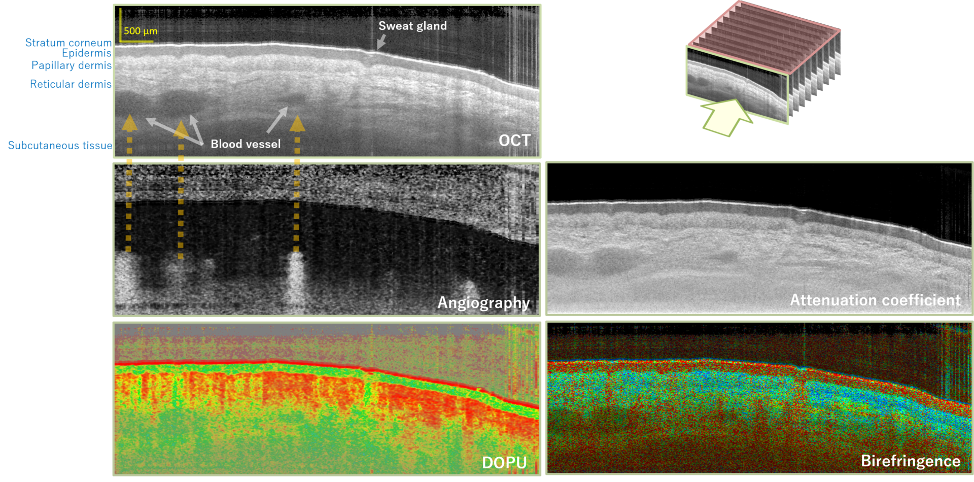
The surface morphology, such as fine wrinkles, texture, and pores, as well as the structure of the blood vessels running underneath, could be confirmed.
In the tomographic image, the layer structure near the surface could be clearly seen, and organs such as sweat glands and blood vessels could also be observed.
The OCT image showed multiple layers, and by referring to the birefringence image, it was possible to clearly see from which layer the structure of collagen had changed.
Also, by referring to the blood flow images, we can consider whether they are blood vessels or other organs.
Thus, by observing several different optical properties, we can notice facts that we could not reach from the individual images alone, and we can analyze the object more accurately.
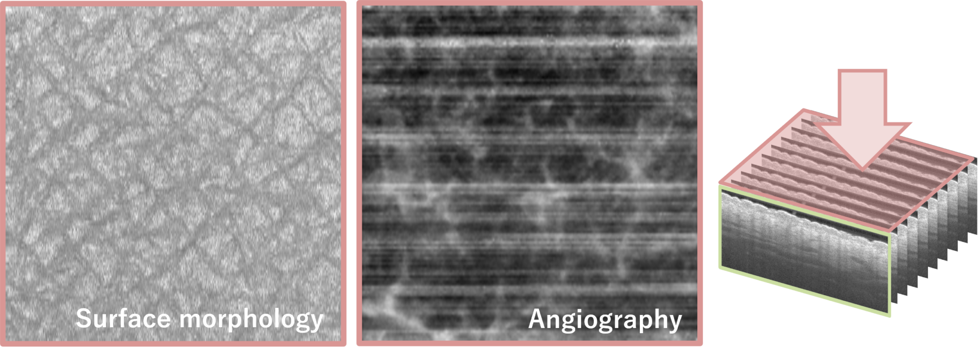
Example of OCT-JM-01 imaging ②
Below is the human forearm measured by OCT-JM-01.
Compared to the surface morphology of the finger, we can see that the grooves (dermal furrows) and the area surrounded by the grooves (dermal papillae) are well aligned. In this state, light is diffusely reflected in a well-balanced manner, and pores are said to be less noticeable.
In addition, when compared to the angiography of the finger, we could clearly see that there are many small blood vessels and that the structure is spread out in a reticular pattern.
Comparing the tomographic images, the thick surface layer visible on the finger was not seen on the forearm (the layer visible on the finger in black on the OCT image, red on the birefringence image, and yellowish green on the degree-of-polarization-uniformity (DOPU) image).
The stratum corneum on the finger is considered to be very thick due to the grasping, and we were able to actually observe this.
The tomographic images also showed that there were many small lumens, not large lumens as seen on the finger.
In this way, it is possible to accurately analyze structures that differ from site to site.
We post new imaging examples on our Facebook page and Twitter from time to time.
OCT-JM-01 specifications
| Lateral resolution | Depth resolution | Imaging depth | Scan range | A-scan rate | B-scan rate | Volume scan time |
| 19 μm | 15 μm (in air) 11 μm (in tissue) | 1.9 mm (depends on the sample) | 6 × 6 × 4 mm3 (you can select 1-12 mm) | 50,000/sec | 80/sec | 6.7 sec |
Functions and performance can be customized according to the measurement target and application. For details, please contact us.
Application example of OCT-JM-01: Kao Corporation ”CT-Skin”
Kao Corporation has developed "CT-Skin," which uses data measured by OCT-JM-01 and applies proprietary algorithms and 3D technology to observe the inside of the skin without cutting.
The link to the news release is here (in Japanese).
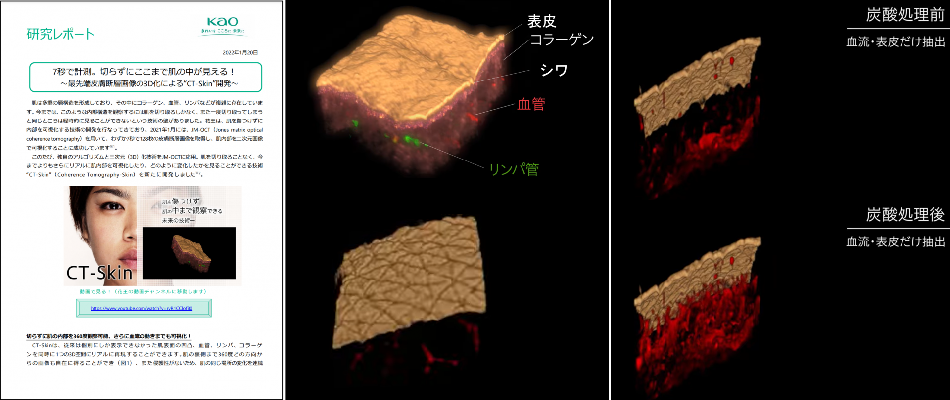
As shown in the video, the skin surface morphology, collagen, blood vessels, and lymphatic vessels were realistically reproduced in one 3-dimensional space.
It also shows how the blood flow changes.
※ Only in Japanese
OCT product specs comparison chart